
Retinal Detachment Treatment in Bangladesh:Symptoms, Surgery and Recovery
Dr Md Maksud Mowla
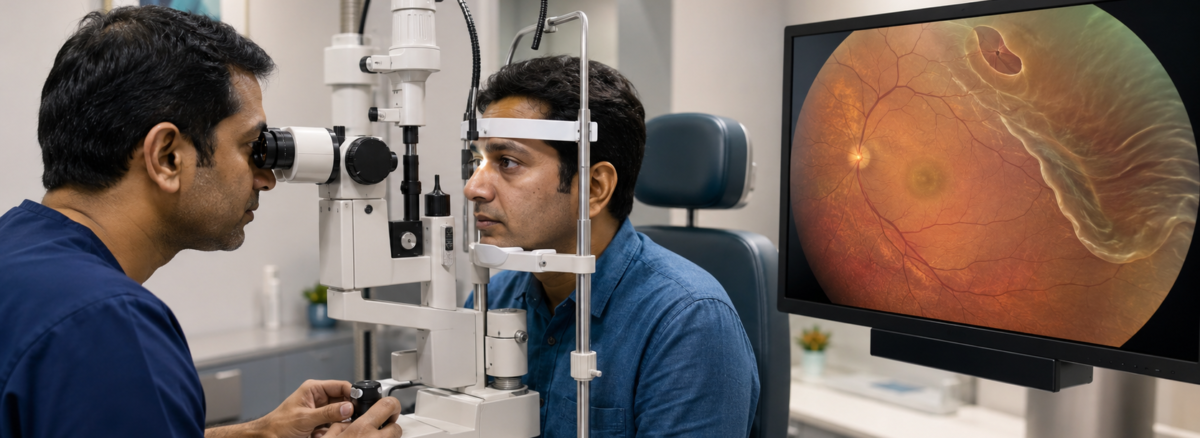
Anyone seeking retinal detachment treatment Dhaka should understand that this condition can be an eye emergency. Sudden flashes, many new floaters or a dark curtain across the vision may indicate that the retina is tearing or separating from the back of the eye.
Retinal detachment is usually painless. Delayed treatment may allow the detachment to spread toward the macula—the part responsible for detailed central vision. Prompt retinal examination may help protect vision, although no treatment can guarantee complete recovery.
What is retinal detachment?
The retina is a thin, light-sensitive tissue lining the back of the eye. It converts light into signals that travel through the optic nerve to the brain.
Retinal detachment occurs when the retina separates from the tissue supporting it. The detached retina cannot function normally, and permanent visual loss can develop if it remains detached.
Rhegmatogenous retinal detachment
This is the most common type. A retinal tear or hole allows fluid from the vitreous cavity to pass underneath the retina.
Tractional retinal detachment
Scar tissue pulls the retina away from the eye wall. This commonly occurs in advanced proliferative diabetic retinopathy.
Exudative retinal detachment
Fluid accumulates beneath the retina without a retinal tear. Possible causes include inflammation, vascular disorders and tumours. Treatment must address the underlying disease.
Warning signs of retinal detachment
Seek urgent retinal care for:
- A sudden increase in floaters
- Flashes of light, particularly in peripheral vision
- A dark curtain, shadow or veil
- Sudden loss of side vision
- Rapidly worsening blurred vision
- Distorted central vision
- Reduced vision after an eye injury
- New symptoms following cataract or retinal surgery
Who is at increased risk?
Important risk factors include:
- High myopia
- Posterior vitreous detachment
- Lattice degeneration
- Previous cataract surgery
- Previous retinal tear or detachment
- Retinal detachment in the fellow eye
- Family history of retinal detachment
- Significant eye trauma
- Proliferative diabetic retinopathy
- Previous intraocular inflammation
- Certain inherited retinal disorders
Retinal detachment treatment in Khulna
Treatment depends on the type, location and extent of detachment; lens status; macular involvement; proliferative vitreoretinopathy; and the surgeon’s assessment.
Laser or cryotherapy for a retinal tear
A retinal tear without extensive detachment may be surrounded with laser photocoagulation or cryotherapy. The resulting adhesion helps prevent fluid from passing through the break.
Laser cannot usually reattach a widely detached retina. It is most effective when the tear is found before significant detachment develops. NEI—Laser and Freeze Treatment
Pneumatic retinopexy
A gas bubble is injected into the eye and the head is positioned so that the bubble closes the retinal break. Laser or cryotherapy seals the break.
This method is suitable only for selected detachments based on the number and position of breaks, retinal visibility and the patient’s ability to maintain positioning. Additional surgery may be required if the retina does not remain attached.
Scleral buckle surgery
A silicone band or segment is placed around the outside of the eye. It indents the eye wall toward the retinal break and reduces vitreoretinal traction.
Scleral buckling may be particularly useful in selected younger phakic patients, detachments without a complete posterior vitreous detachment, certain inferior breaks and some traumatic detachments.
Possible effects include temporary discomfort, refractive change, double vision, infection or buckle-related complications.
Pars plana vitrectomy
During vitrectomy, the surgeon removes the vitreous gel and relieves traction on the retina. Retinal breaks are treated with laser or cryotherapy, and the retina is supported with gas, air or silicone oil.
What determines visual recovery?
Anatomical reattachment and visual recovery are related but not identical. Vision depends on:
- Whether the macula was detached
- Duration of macular detachment
- Preoperative visual acuity
- Retinal ischemia or trauma
- Photoreceptor condition
- Proliferative vitreoretinopathy
- Successful attachment after the first operation
- Cataract or optic-nerve disease
- Postoperative complications
If the macula remains attached, surgery aims to preserve central vision. When the macula has detached, useful improvement is possible, but distortion or reduced contrast may remain.
Vision often improves gradually over weeks or months. Gas, inflammation and changing spectacle power may initially limit vision.
Possible complications
Potential complications of retinal-detachment surgery include:
- Recurrent retinal detachment
- New retinal breaks
- Proliferative vitreoretinopathy
- Cataract progression
- Increased or reduced eye pressure
- Infection
- Bleeding
- Corneal problems
- Double vision
- Retinal displacement or distortion
- Need for additional surgery
- Permanent reduction of vision
The individual risk depends on the original disease and surgical complexity.